Introduction | The Study | Results | My Conclusion
Lewis AM, Layzer R, et al. “Magnetic Resonance Neurography in Extraspinal Sciatica” Arch Neurol. 2006;63:1469-1472
When it comes to low back pain with associated radiating lower limb pain (sciatica), lumbar disc herniation and/or stenosis are the usual suspects with regard to causation. However, the clinical picture gets more complicated when the former and latter are not present on imaging, yet the patient suffers debilitating sciatica. What could be the cause?
Besides other possible nonpathological causes (like discogenic referred pain or instability from a spondylolysis), abnormality within the piriformis and/or gemellus musculature have been proposed as a possible cause of the relatively rare extraspinal sciatica. More specifically, since the sciatic nerve passes between the piriformis and gemellus musculature, it has been speculated that hypertrophy in those muscles (as well as involvement of a adjacent fibrous band of connective tissue ) may compress the sciatic nerve in the pelvis, which in turn results in the patient's sciatica.
The diagnosis, however, is not easy to make. This is because the sensitivity and specificity of examination and orthopedic findings is quite low for the condition. (In other words, doctors can't really tell if you have it or not from their exam). Furthermore, MRIs sensitivity and specificity is also not very good. The good news, is that we have a new tool which ostensibly boasts a higher sensitivity and specificity for making the diagnosis!
In 2006, Kaiser’s Lewis et al, published the results of their retrospective investigation into the diagnostic value of MR neurography for making the diagnosis of extraspinal sciatica – i.e. piriformis syndrome.
The Study:
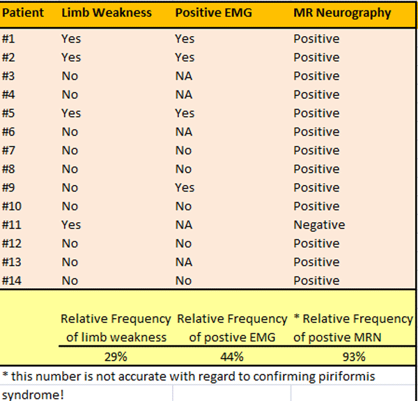
The cohort (group of patients) was created from a retrospective (done after the fact) review of medical records of 14 patients that suffered unexplained unilateral sciatica – i.e., these patients had no disc herniation, stenosis or any other obvious cause of the sciatica. All of these patients had previously undergone MR neurography as part of a further diagnostic workup. The subsequent images from MR neurography were interpreted by one of the researchers, all of whom specialized in reading MR neurograms.
Results
Unfortunately, this was not a well written paper and interpreting the results was challenging to say the least. The authors reported that in 12 of patients (86%), MR neurography demonstrated signs of inflammation and other pathological change within the sciatic nerve.
With regard to the portion of the sciatic nerve adjacent to the piriformis muscle, only 8 of them (57%) had signs of inflammation.
Study Confounders
There were plenty of things that damaged this study. Let's take a look at them.
- the MR neurography in one patient (7%) demonstrated inflammation in the sciatic nerve on both sides: the sciatic-affected leg, as well as the normal leg.
- another patient had a completely normal MR neurography, yet he had unexplained sciatica. To make things more confounding, this patient ultimately underwent a successful piriformis release-- the patient's sciatica got better from releasing the sciatic nerve from entrapment within the piriformis muscle (so MR neurography completely missed the boat here).
- another patient had positive MR neurography on the ipsilateral side, but the inflammation in the sciatic nerve was not at the level of the piriformis muscle – it was all the way down at the level of the ischial tuberosity (far away from the piriformis muscle).s
- the study was way underpowered! We need at least 50 patients in order for the study to be worth anything.
More Results
Conclusion/Comments
This was a very small retrospective study aimed at looking inside the sciatic nerve in patients with unexplained sciatica. Of the 14 patients, 12 (86%) had focal inflammation within the ipsilateral sciatic nerve. Only 8 of the 14 (57%) showed signs of piriformis syndrome as confirmed by MR neurography.
There were forming confounders in this tiny study that really suggests MRN is not a useful test for making the diagnosis of piriformis syndrome. (See above)
Obviously, a much larger study needs to be done to determine with regard to the usefulness of MRN for the diagnosis of piriformis syndrome.